Drusen In The Eyes: What Are They And How Do They Affect Sight
Drusen in the eyes are deposits of waste that accumulate because the body is not able to eliminate them. Depending on where these accumulations appear, they are called macular drusen or optic nerve drusen .
The truth is that they are a frequent phenomenon that is associated with age. The problem is that, if they are large or numerous, vision can be affected. Especially the central. That is why in this article we explain everything you need to know about this situation.
What are drusen in the eyes?
Drusen in the eyes, according to an article published in the Archives of the Spanish Ophthalmology Society, are accumulations of amino acids, iron, calcium and ribonucleic acids. The fact that they contain calcium makes them more opaque and prevents the passage of light towards the retina.
They are usually yellowish in color. Its pathogenesis is not well understood, but it is known that macular drusen are more common as we age. However, those of the optic nerve do not seem to be associated with this fact.
Even optic nerve drusen (DNO) can appear in children. According to a study published in the Annals of Pediatrics, in infants they do not usually cause symptoms, but sometimes they cause a decrease in visual acuity.
Why are they generated?
As we have mentioned, it is not very well understood why drusen occur in the eyes. According to the American Academy of Ophthalmology , most cases of macular drusen are age-related.
That is why they usually appear in people over sixty years of age. In these cases, when the deposits reach a large size, they are associated with the macular degeneration of aging.
In addition, certain factors have been detected that can lead to the appearance of drusen in the eyes. It seems that they are more frequent in the Caucasian race. Similarly , high blood cholesterol levels or tobacco can act as a risk factor. Lastly, having a family history also increases the likelihood.
Types of drusen in the eyes
Drusen in the eyes can be classified into different types, according to what age they appear, their evolution and the risks they entail. A study published in the Spanish Retina and Vitreous Society places hard drusen first. These are usually located between the retinal pigment epithelium and Bruch’s membrane. They are considered a normal sign of aging.
Soft drusen are located in the same place as hard drusen. However, they are associated with an increased risk of developing eye problems. That is why it is recommended to do a follow-up every six months.
Symptoms and diagnosis of drusen in the eyes
Drusen in the eyes may or may not produce symptoms. This depends, above all, on how many there are and the size they reach. When there are signs, central vision is often lost or flashing lights appear in the field of vision.
Some people experience blurred vision. There may also be darkening, black spots in the field of view, or problems going from a bright environment to a darker one.
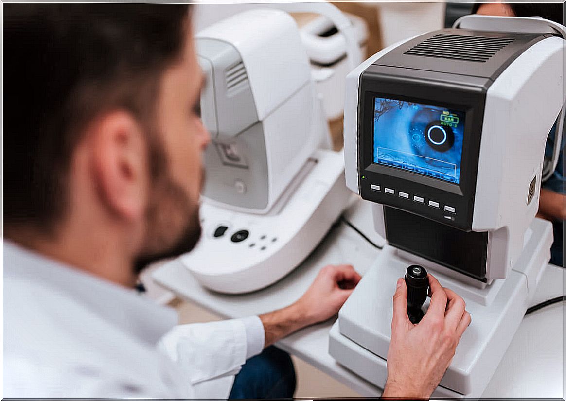
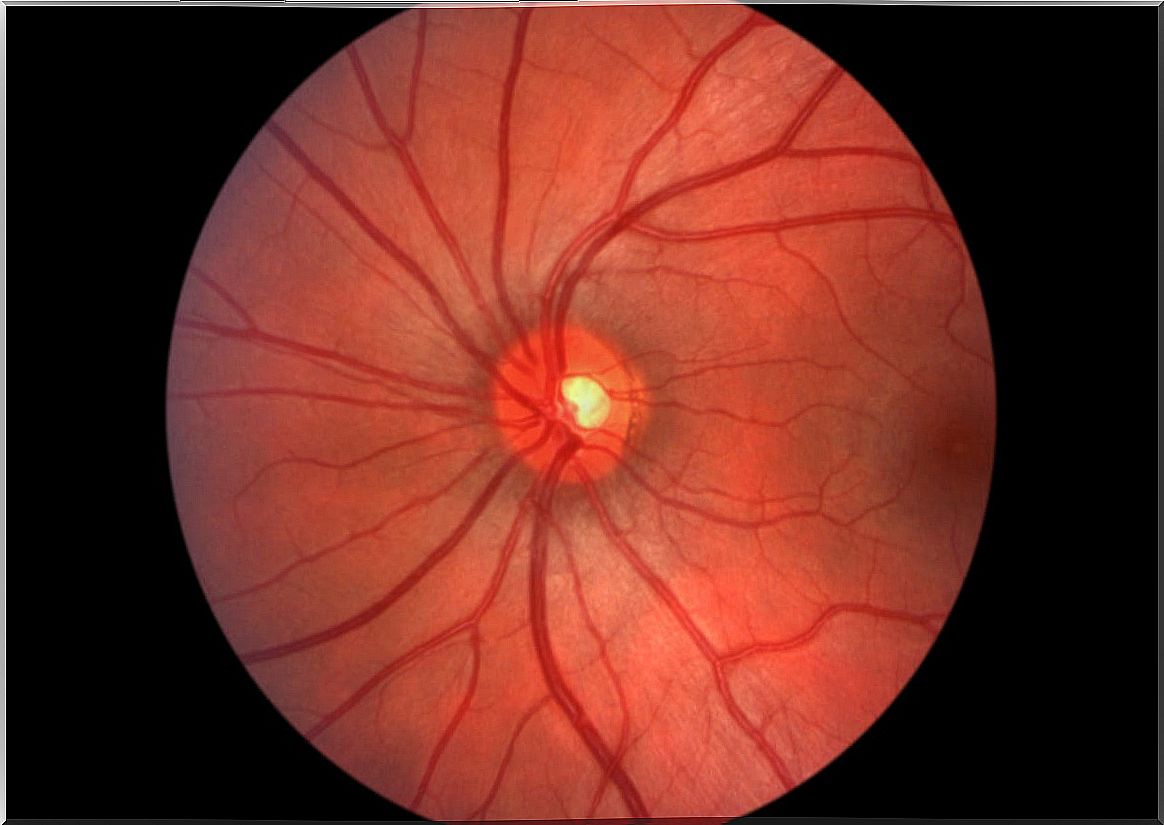
To diagnose them, in addition to the clinic, it is essential to perform an eye examination. What is done is to observe the fundus to be able to check the state of the area around the retina.
Other more specific tests are optical coherence tomography and fluorescein angiography. The first is a non-invasive technique that allows the macula and optic nerve to be explored. Light waves are used to obtain cross-sectional images of the retina.
Fluorescein angiography involves injecting a contrast agent (fluorescein) into a vein. This contrast diffuses through the body and reaches the blood vessels of the eyes. Images are taken of them using a special camera. This is how you can see if there is any injury to them.
Another test that is also commonly used is the Amsler grid test. This allows to evaluate the central vision. When it is altered, it is oriented towards a possible macular problem.
Complications
As with the symptoms, the complications of drusen in the eyes will depend, above all, on the number and size of them. That is, the more there are and the larger the size, the more frequent alterations in vision appear.
One of the first complications is retinal tissue atrophy. This impairs central vision. But the most prominent disorder is macular degeneration. It is a degenerative disease that, when it occurs in the macula, also alters the central visual field. Similarly, the ability to observe details is lost.
Drusen of the optic nerve, as explained in a study published in the Archives of the Spanish Ophthalmology Society, can lead to severe campimetric defects.
Can drusen in the eyes be prevented or treated?
As we have seen, a large part of the drusen in the eyes is associated with aging. However, we also commented at the beginning that certain factors, such as tobacco or cholesterol, can lead to its appearance.
Therefore, leading a healthy life, with a balanced diet and including daily exercise is key. In this way, both cholesterol and glucose levels in the blood are improved, which protects the retina. Sunglasses are also recommended to prevent damage from UV radiation.
The treatment itself depends on the type of drusen and its progression. For example, in the case of soft ones, it is recommended to start a treatment to avoid macular degeneration. The most widely used drugs are antiangiogenic drugs that prevent the formation of new vessels around the retina.
This is beneficial, as the vessels that usually form are weaker and more fragile. That is why it is common for them to break or for fluid to pass through them, causing small hemorrhages or edema around the macula.
Cuticular and reticular drusen should also be treated with antiangiogens. On the contrary, in the case of the hard ones, the only thing that is recommended is a routine control, since no treatment seems to be effective.
Remember that you must go to the ophthalmologist
The most important thing to keep in mind about drusen in the eyes is that, in order to detect and treat them before they cause serious vision disturbances, it is essential to go to the ophthalmologist.
Many of them appear with age, but can be treated to prevent them from progressing. Therefore, if drusen are detected in the eyes, it is essential to go to all the recommended controls. In general, it is sufficient to go every six months or a year.